What is facet pain syndrome?

Facet joint pain is one of the main causes of lumbalgia, accounting for up to 40% of cases of pain in the lumbar spine. This pain originates in the facet joints, which are located in the posterior area of the vertebrae.  

Facet joints are synovial joints that have hyaline cartilage, synovial membrane, a fibrous joint capsule, and joint space. Like other joints, with the passage of time and the loads they are subjected to, they are prone to a process of degeneration.  

Causes of facet joint pain

The degeneration of the synovial joints is produced by the sum of multiple genetic and environmental factors, and in most patients, there is not a single determining cause. In some cases, there can be a known primary cause, such as injury or congenital malformation, among others, with osteoarthritis being secondary to these.   

Thus, any degenerative process of the joint or its elements can produce facet joint pain. The main cause is osteoarthritis, although other pathologies such as tears of capsular ligaments, synovial cysts and injury or degeneration of articular cartilage can also cause this disease. 

Alteration of elements of the anterior area of ​​the vertebra, such as the processes of disc degeneration, can affect the posterior balance or cause spinal instability, which will result in increased loads on the joints and greater wear on their components. 

Symptoms of facet joint pain

In the initial stages of joint degeneration, the patients do not notice any symptoms, so by the time the patient begins to notice facet joint pain, these joints have already undergone many changes.  

When symptoms become noticeable, they usually include pain, stiffness, and restricted movement. Stiffness increases after periods of reduced joint mobility, such as sitting for a time and then getting up from the chair, or getting up from bed in the morning. In addition, the pain increases in movements in which the joint space is reduced or the pressure on them is increased, such as when standing or carrying something heavy

When the pain is in the area of the lumbar spine, as is most frequent, the clinical picture includes pain in the lumbar or lumbosacral region, which, in some cases, radiates to the buttocks and the back of the legs down to the knees. When the cervical area is involved, the patient suffers neck pain that radiates to the nape of the neck, the shoulder girdle and proximal area of ​​the upper limbs, without root pattern of the spinal nerves. 

Diagnosis of facet pain

The diagnosis of facet pain is not easy. There are several pathologies of the spine that share common symptoms and signs, and the patient can have more than one joint problem, so a complete study is required

Diagnosis begins with knowing the patient’s symptoms, which, as mentioned above, can include pain, stiffness and limited mobility. During the physical examination, it may be discovered that palpating the facets causes pain. In addition, the pain increases with spinal extension or extension-rotation maneuvers. 

Anteroposterior x-rays can be used as imaging tests, where sclerosis and increased joint volume can be observed, generally visible in cases where the degeneration is advanced. In the oblique view, a decrease in joint space can be observed, including osteophytes, which are both signs of joint osteoarthritis. 

Nowadays, the x-rays are usually supplemented with other imaging tests such as a CT scan or magnetic resonance imaging, which can show in greater detail if there are structural alterations that would explain the patient’s symptoms, from arthritic involvement of the facets to adjacent joint lesions, such as cysts. 

The pain does not always match the information shown in the scans and x-rays, so there are patients with symptoms and yet their imaging tests show nothing to explain them. In these cases, and in cases with multifactorial pain where it is difficult to know how much of the pain is due to the facets, the performance of clinical-diagnostic infiltrations is indicated. This procedure is guided by fluoroscope, with local anesthetic and corticosteroids. For about 60-80% of patients with facet pain syndrome, such facet joint injection yields improvement. 

Treatments - facet joint pain syndrome

Initially we begin with conservative treatment for pain management. The use of NSAIDs is recommended, and the drug treatment for pain relief can be modified according to the patient’s indications on the WHO pain scale. Specialized physical therapy guided by a professional, is also recommended, to perform stretching and muscle strengthening exercises.  

Intermediate and surgical treatment

In cases where the patient’s symptoms do not improve with conservative treatments, minimally invasive interventionist techniques are used. The most frequently chosen option is thermal radiofrequency ablation (rhizolysis), which consists of performing a controlled lesion to the medial branch of the posterior root of the corresponding spinal nerve. This controlled nerve injury is provoked by a local increase in the vibration of the particles surrounding the active tip of the cannula. Between 60-90% of patients experience significant improvement in pain.  

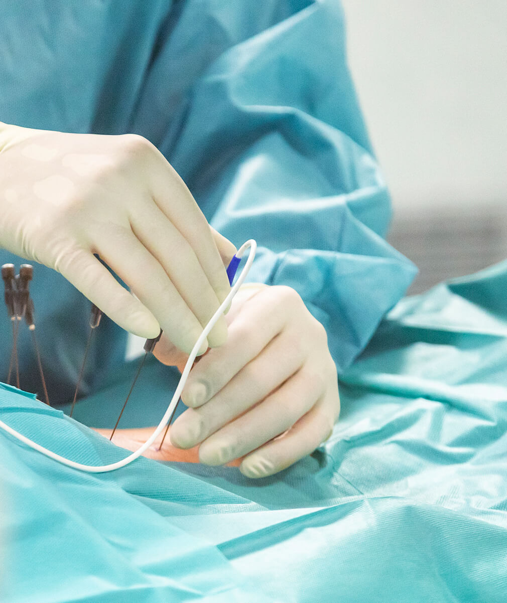
The minimally invasive technique of joint injections is performed in an operating room, with light sedation that allows patient collaboration. The entire procedure is monitored with a fluoroscope. A needle with a sensor in its tip is inserted into each of the affected joint or joints.  

First, a sensory check is performed (50 Hz), followed by a motor check (2 Hz), in which we do not want to see root stimulation, although fasciculations may appear. Once the safety of the procedure has been verified, the radiofrequency is started. There are two types, the conventional one, which uses heat at 80ºC for 60-80 seconds, or the pulsed one, which is usually carried out for 120 seconds and reaches temperatures of 40-42ºC.     

This minimally invasive technique can be performed on the entire spine from the cervical area (starting at C2) to the lumbar and sacral area. In addition, it can be unilateral or bilateral, depending on the patient's situation. Most patients experience immediate improvement, although this may not be noticed until up to 4 weeks after the procedure. 

It is considered a safe technique, with a low rate of complications. Despite this, there are patients who experience pain or discomfort in the operated area, and there have even been cases of nerve pain in the form of a burning sensation or dysesthesia, which improves within 4-8 weeks afterwards. 

It should be noted that the neural blockage achieved lasts for a given period of time, and the pain may reappear again. It is difficult to predict how long the pain relief from this treatment will last for each patient, since it can vary from 6 months to several years. It is good to know that if the facet joint pain reappears, the facet joint injections can be performed again

Who are the doctors at Instituto Clavel who treat facet pain syndrome?

Facet joint pain can be treated by any specialist on the team

Sources

  • Haralson R, Zuckerman J. Prevalence, health care expenditures, and orthopedic surgery workforce for musculoskeletal conditions. JAMA 2009; 302 (14): 1586 - 1587
  • L. Kalichman, L. Li, D. Kim, et al. Facet Joint Osteoarthritis and Low Back Pain in the Community - Based Population. Spine, 33 (2008), pp. 2560-2565
  • F. Andrés Chahín, C. Carlos Valenzuela. Evaluación y manejo del dolor lumbar de origen facetario. Rev. Med. Clin. Condes 2014; 25(5) 776-779
  • 4.J. Cavanaugh, et al. Pain Generation in Lumbar and Cervical Facet Joints. J Bone Joint Surg Am, 88A (2006), pp. 63-67
  • 5.J. Schofferman, D. Mazanec. Evidence - informed management of chronic low back pain with opioid analgesics. Spine J J, 8 (2008), 185-194
  • 6. Niemisto L, Kalso E, Malmivaara A, Seitsalo S, Hurri H. Radiofre-quency denervation for neck and back pain. EBM Reviews, Co-chrane Database of Systematic Reviews. 2007;1
  • 7. M.L. Franco. Bloqueos diagnóstico-terapéuticos de carillas articulares cervicales. Rev Soc Esp Dolor. 2009;16(2):116-121
  • 8. E.J. Carragee, El Hurwitz, I. Cheng, et al. Treatment of Neck Pain: Injections and surgical interventions. Results of the Bone and Joint Decade 2000–2010. Task Force on Neck Pain and Its Associated Disorders. Spine, 33 (2010), pp. S153-S169

Want to know more?
Tell us about your case!

Contact us so that we can give you a personalized assessment.

Share on: