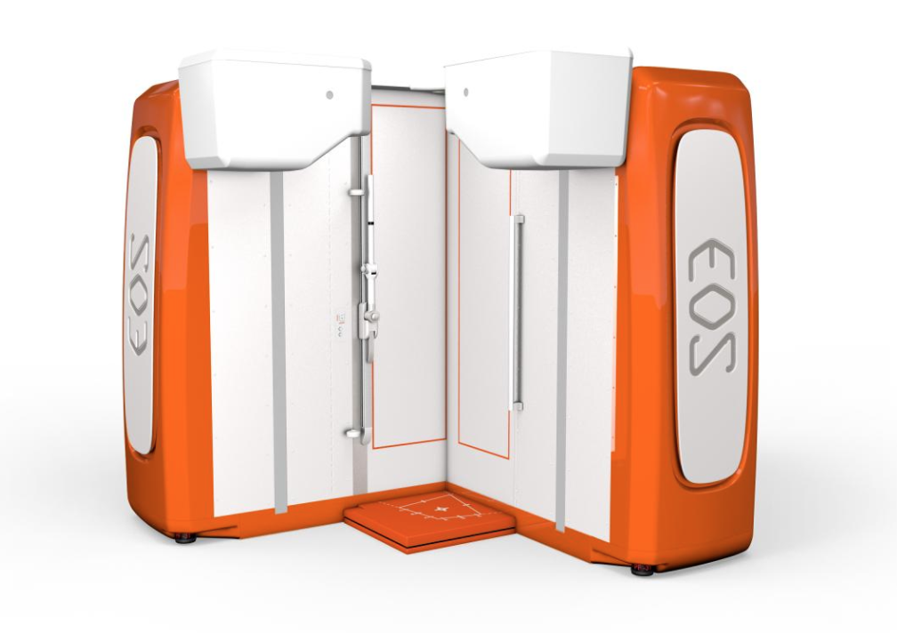
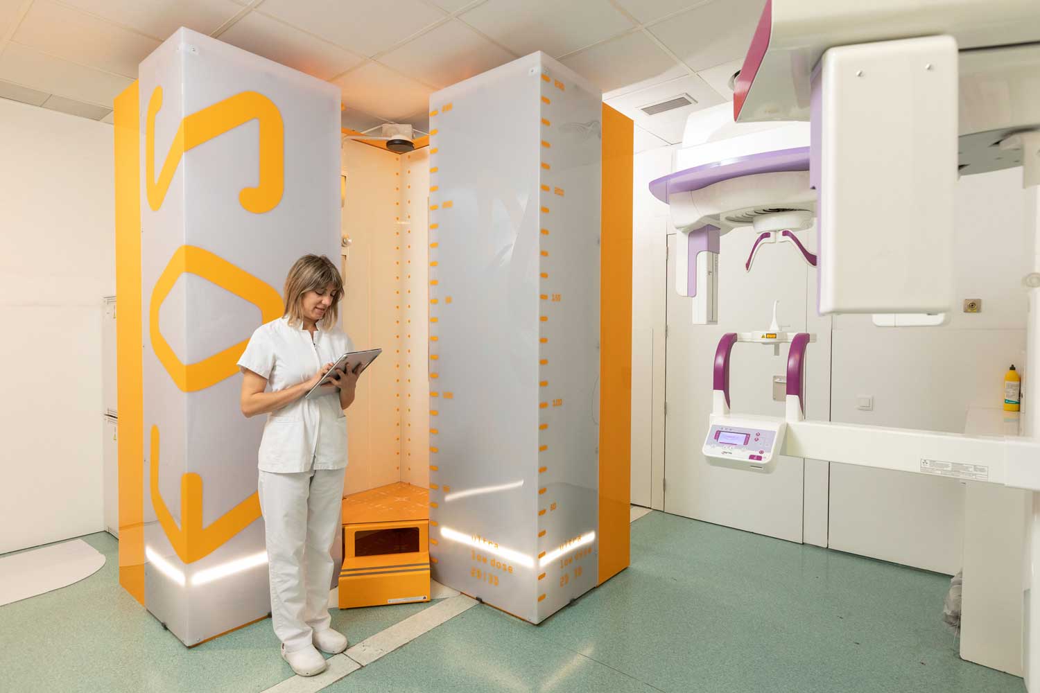
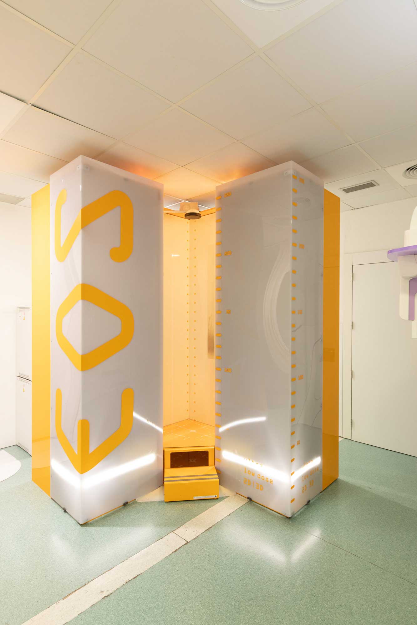
Every person is unique and each person’s spine is different. The form of the spine changes over time, as we grow and age, and can also change due to an illness affecting this part of the body. This is why making a precise diagnosis is essential to apply the appropriate treatment. At Instituto Clavel we have the EOS technology to achieve this.
Studies over the past 15 years have taught us that there are different types of healthy spines, with different shapes and morphologies, and depending on the type, they require radically different treatment and planning. The different forms of healthy spine described in the Roussouly classification are determined by a set of spinopelvic parameters, which defined by the measurement of angles and distances between the head, spine, pelvis and legs.